Goldenhar Syndrome – Introduction and Basic Points

What You Need To Know About Goldenhar Syndrome

The rare congenital condition known as Goldenhar syndrome is characterized by a variety of abnormal developments in association to the spine, ear and eye. Also known as OAV (Oculo-Auriculo-Verterbral Spectrum), this congenital condition was documented first by Maurice Goldenhar in 1952, a general practitioner and ophthalmologist. It is believed to affect 1 in every 3000 to 5000 births.

Babies that are born with Goldenhar syndrome will typically have totally absent or partially formed ears, spinal deformities like scoliosis and benign growths over the eyes. Goldenhar syndrome can also affect the child’s facial structure or organs such as the nervous system, lungs, kidney and heart. In the majority of cases, deformities will usually occur on 1 side of the child’s body.

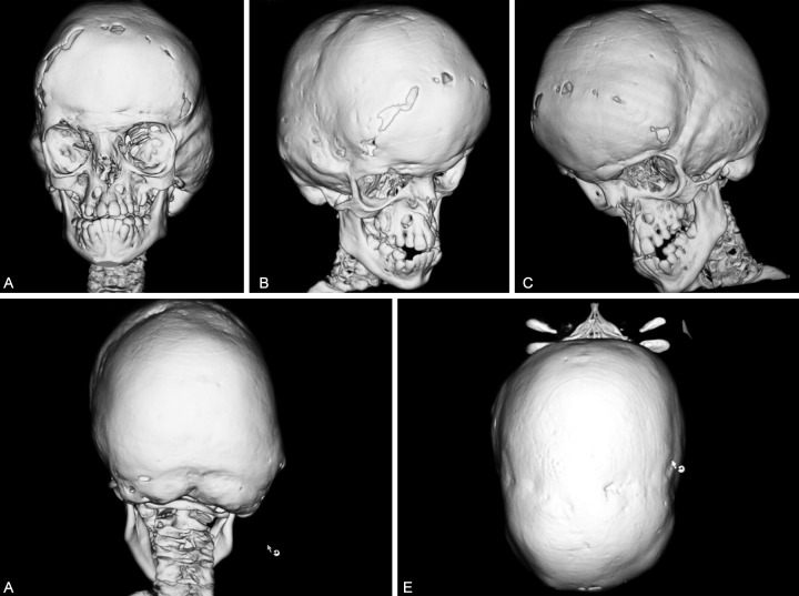
One factor of Goldenhar syndrome has to do with hemifacial microsomia. This means the cheekbones and the jaw on 1 side of the child’s face will be undeveloped. The underdevelopment, combined with ear and eye anomalies, results in distinctive types of facial features for the children that have Goldenhar syndrome.

Rib cage and spine deformities are another common aspect of Goldenhar syndrome. In certain cases, the ribs or vertebrae inside the spine do not fully form, are fused abnormally or they are missing. About 50% of children that have Goldenhar syndrome will present a type of congenital-scoliosis. The spine anomalies result in pulmonary disorders and incomplete growth.

Symptoms And Signs

The symptoms associated with Goldenhar syndrome will vary, but can include the following features:

• Spinal abnormalities that lead to Kyphosis, scoliosis or both

• Cardiac defects

• Abnormal rib structure that includes fused or missing ribs, this leads to decreased lung function, thoracic insufficiency and poor growth

• Hearing loss, that typically occurs in 1 ear

• A variety of craniofacial disorders that include:

Cleft palate or cleft lip
– Hemifacial microsomia, which involves tissues on both sides or one side of the child’s face that is underdeveloped, especially in areas that affect the jaw, mouth and ear areas
– A mouth that is wide, with one side that is slightly higher compared to the other
– Benign growths or cysts over or around the eyes known as ocular-dermoid cysts
– Totally absent or partially formed ears known as microtia

• Central-nervous-system defects

• Respiratory issues

• Urogenital and kidney issues

Treatment

Due to the fact that Goldenhar syndrome has the potential to affect a number of the body systems, the treatments for this condition will vary. In certain cases, monitoring with care is all that will be necessary. For other cases, surgery will be required in order to address certain aspects of this condition.

A variety of complications in association to Goldenhar syndrome will be evident when the baby is born and are treatable while the child is still young. Some of these examples include craniofacial anomalies, hand disorders and club feet.

Other types of complications of Goldenhar syndrome can only start to show up or become problematic as the child grows. These issues usually include joint disorders, dislocated hips and spinal deformities.

Each child’s condition will be different, which means a treatment plan will be determined in regards to a case-by-case basis. Treatments can include non-surgical options like physical therapy and bracing, or surgical treatments such as implanting and then expanding growing-rods to stabilize the child’s spine as they grow or spinal fusion.

Follow-Up Care

A child will Goldenhar syndrome must be monitored by orthopaedic physicians into their adult years. If the child has undergone spinal-fusion surgery, they will have appointments scheduled with an orthopaedic surgeon a week after the surgery and then 3 to 6 months post-surgery.

girl smiling

Understanding Malocclusion of the Teeth

Malocclusion of the teeth is a severe misalignment that can lead to more severe dental conditions. This condition has also been called overbite, open bite, under-bite, cross bite, or crowded teeth.

girl smiling

If teeth do not properly connect, or if they’re misaligned, they will not be able to perform the vital functions they are designed for. The best way to protect your vital functions like bone structure and digestive health, it is important to be conscious of the role the teeth play.

Occlusion can be best described as the alignment of the teeth. The optimal layout include neatly spaced teeth that are not twisted or turned in anyway. The teeth of the upper jaw neatly fold over the teeth of the lower jaw and the pointed ridges in the molars interlock perfectly with those above.

Malocclusion is the term given to deviations from this optimal form. The deviations can vary to a great degree but the point is that all misalignment and occlusion will affect dental function in some way. Proper alignment of the upper teeth keeps the cheeks and lips from being bitten, while the proper alignment of the lower teeth protects the tongue from being bitten.

1. What Causes Malocclusion?

Malocclusion is a typically inherited condition, which means that the genes of the parents will pass on the malocclusion to children.

Then there are other specific habits and conditions that can contribute to the misalignment of the teeth. These include:

–Prolonged feeding from bottles during early childhood.
— tumors of the jaw and mouth
Cleft Palate or lip
–abnormally shaped or impacted teeth.
–The use of pacifiers during infancy and before the age of 3.
–injuries that result in the misalignment of the jaw
–improper dental care that can include ill-fitting braces and defective crown, fillings or bridges.
–airway obstructions that can cause mouth breathing, like allergies adenoids etc.

2. What Are the Symptoms of a Malocclusion?

Depending on the severity of the condition, the symptoms of malocclusion can be very subtle or very pronounced. Some of these symptoms include.

— speech problems, including the development of a lisp
— discomfort when chewing or biting
— improper alignment of the teeth
— breathing through the mouth rather than the nose
— alteration in the appearance of the face
— frequent biting of the inner cheeks or tongue

3. Diagnosing and Classifying Malocclusion

Malocclusion is classified in different ways depending on the type of deviation and its severity. Your dentist will conduct routine dental examinations and take x rays to determine which classification your malocclusion can be.

The three categories of malocclusion include.

Class 1– the most common type of malocclusion occurs when the teeth of the upper jaw overlap the teeth of the lower jaw. The bite is comparatively normal and the the deviation is slight.

Class 2 — this category of malocclusion covers all severe overbites. This condition also has the name retrognathism (or retrognathia). Referring to the severe overbite of the upper law and overlap of the lower teeth.

Class 3 — this type of malocclusion occurs when there is a severe under-bite present. Also called prognathism, and refers to protrusion of the lower jaw, this can cause the teeth of the lower jaw to overlap those of the upper jaw.

4. How Is a Malocclusion of the Teeth Treated?

The vast majority of malocclusion conditions will require no treatment. If the malocclusion is severe enough to affect your dental function, your dentist may refer you to a qualified orthodontist. Your orthodontist will recommend any one or a combination of the following treatments.:

— reshaping, bonding, or capping of teeth
— wires or plates to stabilize the jaw bone
— braces to correct the position of the teeth
— removal of teeth to correct overcrowding
— surgery to reshape or shorten the jaw

Some of the more common conditions that can result from treatment of this type include:

— irritation of the mouth due to the presence of an appliance.
–pain and severe discomfort
–difficulty speaking and chewing after the procedure.
–increased possibility of tooth decay.

5. How Can Malocclusion Be Prevented?

Because the majority of malocclusion cases are hereditary, it is hard to prevent them. Nevertheless, parents who are aware of this condition in their bloodline should consider taking action and preventing pacifier and bottle feeding from aggravating the condition. Early detection is also an important way to begin administering adjustments in formative years while the full effects of the malocclusion have no yet been consolidated.

Early treatment of the conditions and deviations will make treatment time considerably shorter.

The costs of treatment will also be much cheaper.

Nevertheless, no matter your age, treatment for this condition has invariably improved the quality of life of the individual.

cleft-lip

Common Orofacial Cleft Conditions

Cleft palate and cleft lip known as orofacial clefts are a common type of birth defect which occurs when an infant’s mouth or lip does not develop correctly during pregnancy. People who have these conditions will often have to undergo a number of surgeries along with other types of medical procedures.

Even though the exact causes associated with orofacial cleft conditions is unknown, the affected children usually have a close or distant relative that had an orofacial cleft. This is a suggestion that genes can be a deciding factor as to why some people contract this condition.

Associated Conditions With Orofacial Cleft

In many cases when a baby is born presenting a cleft palate or cleft lip there are chances that they have other medical conditions. The associated conditions often include 22q11 Deletion Syndrome, Stickler Syndrome and PRS (Pierre Robin Sequence). The pregnant mothers that have received a diagnosis for their unborn child with a orofacial cleft condition, will in most cases be offered with additional tests as these babies have a higher chance of having other syndromes when compared to a baby without orofacial clefts. Some mothers undergo amniocentesis tests to try and detect syndromes that have a greater impact like Down’s and Edwards syndrome.

One of the more common conditions often associated with cleft is PRS (Pierre-Robin Sequence). This condition is typically detected once the baby is born and the baby will have a very small chin. These infants will usually have cleft plates as their tongues are not able to fall into position correctly while the infant is developing. This cleft is typically U-shaped and wide and will have an effect on the hard-palate and the soft palate.

Babies that are born with PRS may have breathing difficulties and CNS (Clinical Nurse Specialists) will advise the mother on an “individual basis”. However, they often suggest that the infant feed and sleep on “their” sides for a number of weeks after the birth. Some babies with this condition will require assistance with their airways and in some cases a naso-gastric tube will be used to deliver food directly into the stomach and to avoid the risks of milk that goes down incorrectly during feeding.

Stickler Syndrome is another condition associated with a cleft lip or palate. This is a far rarer condition and in some cases will go un-diagnosed. SS (Stickler Syndrome) is recognized as a type of genetic condition which is typically passed from one of the parents onto the child. This condition is common in both girls and boys. Having this condition decreases the amounts of connective tissue and collagen that support the organs or cover for the bone ends inside each joint. Collagen also assist with the normal functioning of the eyes, so babies that are born with this syndrome will typically need frequent check-ups at eye-hospitals. Stickler Syndrome is also linked with the heart murmurs like “mitral valve prolapse”, and also will require regular visits to a Cardiologist or Pediatric heart doctor.

22q11 Deletion Syndrome that also goes by the name of Conotruncal Anomaly Face Syndrome, Shprintzen Syndrome and DiGeorge Syndrome. This syndrome happens due to a fault that occurs in chromosome 22 which usually results in unusual or characteristic facial features, abnormalities of the heart and palate issues such as an isolated cleft palates or sub-mucous plates whereby a layer of thin skin covers the cleft. Other symptoms can include the soft plate that may appear normal but causes issues with feeding or speech, lowered immunities to infections, blood-calcium levels that are low. The babies that are born with 22q11 Deletion Syndrome can have one or all of the above mentioned conditions.

Crouzon-skull-scan

Crouzon Syndrome – Symptoms and Background

What You Need To Know About Crouzon Syndrome

Crouzon syndrome is classified as one of the rare inherited disorders whereby a variety of the sutures (flexible seams) in the skull of a baby fuses or turn into bone too early. This early fusion is a hallmark for a variety of conditions named craniosynostoses.

In a normal baby, the flexible seams (sutures) in the infant’s skull will remain open in order to allow the normal growth of the brain. When the sutures close up too early, the face and the skull can or will become misshapen due to the growing brain. The signs associated with Crouzon syndrome usually begin within the first months of the baby’s life. This continues and progresses until the child’s 2nd or 3rd birthday.

This syndrome affects around 5% of all the babies that have craniosynostosis. Louis E. O. Crouzon a French neurologist first described the condition in an earlier part of the 20th century.

Crouzon-skull-scan

Symptoms

The babies that have Crouzon syndrome can present the following symptoms:

• Bulging eyeballs

• Enlarged forehead

• Eyes that will point in 2 different directions

• A narrow and long head or a wide and short head

• Eyes that are spaced widely apart

• Strabismus, which means crossed eyes

• Eyelids which slant downwards

• Vision loss

• Beak-like and curved nose

• Flattened cheeks

• A lower jaw that protrudes

• Short upper lips

• Poorly developed and small upper jaw

• Crowded teeth

• Hearing loss

• Cleft lip (opening in the lip), or cleft palate (opening in the roof of the mouth)

• Mismatched bite

The above mentioned symptoms can be more drastic in certain babies when compared to others. A very small percentage of Crouzon syndrome children will also present a skin condition known as acanthosis nigricans. The condition results in thick, rough and dark patches on the skin. They develop in the folds such as the groin, neck, armpits and behind the child’s knees.

Life Expectancy

People who have Crouzon syndrome usually live a normal life-expectancy and the majority of the children diagnosed with the condition will be unaffected intellectually. However, the condition can result in altering the face shape which can result in hearing and vision issues.

Causes

Crouzon Syndrome occurs due to changes that are known as mutations in 1 out of the 4 FGFR genes. In most cases it will affect the FGFR2 gene and in rare cases it will affect the FGFR3 gene.

The genes perform the role of carrying out instructions in regards to making proteins which direct the functions of the body. A mutation is able to affect the functions that the specific type of protein has.

The FGFR2 codes for the protein known as “fibroblast growth factor receptor 2.” As the fetus is developing inside the womb, this is the protein which signal the bone cells to develop. The mutations of this specific gene increase these signals, which is what increases the bone development which is what results in the skull fusing way too early.

The baby that inherits this syndrome only needs to take on one copy of this gene mutation derived from either parent in order to contract Crouzon syndrome. If you happened to have the condition, each child that you have will have a 50% chance in regards to inheriting it. This type of inheritance-pattern is known as “autosomal dominant.”

In around 25 to 50% of the individuals with Crouzon syndrome, gene mutation occurs spontaneously. When this occurs, the baby did not need a parent who has the syndrome in order to contract this disorder.

Complications Associated With Crouzon Syndrome

The typical complications that have been reported with Crouzon syndrome include:

• Vision loss

• Hearing loss

• Hydrocephalus (fluid build-up inside the brain)

• Breathing problems or sleep apnea

• Exposure keratitis (inflammation in the front of the eyes), or exposure conjunctivitis (inflammation in the membrane lining the whites of the eyes).

Treatment

Children that have been diagnosed with a mild form of Crouzon syndrome might not require treatment. The children who have been diagnosed with severe Crouzon syndrome should be seeing a craniofacial specialist who is doctors that are experienced and treat the disorders associated with the face and the skull.

In the cases that are more severe specialized doctors are able to conduct careful surgery that opens up sutures, which allows the brain the space to grow normally. After such a surgery the child will be required to wear a specialized helmet. This helmet assists in reshaping the skull.

Surgery is usually required for the following reasons:

• To straighten or correct crooked teeth

• Relieve the pressure that is building up in the skull

• Correction of eye issues

• Corrections for malformed jaws

• To correct cleft palates or cleft lips

Children that have hearing issues caused by Crouzon syndrome are able to use hearing aids in order to amplify sounds. Children diagnosed with the condition can also require language or speech therapy or both.


Here’s an encouraging story. A little change can go a long way …

Say hello to Charlie


What Is Involved In The Diagnosis Of Crouzon Syndrome

The diagnosis of Crouzon syndrome usually begins with a doctor examining the face and the skull shape. Doctors also make use of imaging tests that include MRI scans (magnetic resonance imaging), CT (computed tomography) and X-rays to locate an increase in pressure inside the skull or fused sutures. Tests to locate a mutation of the FGFR2 gene are also used.